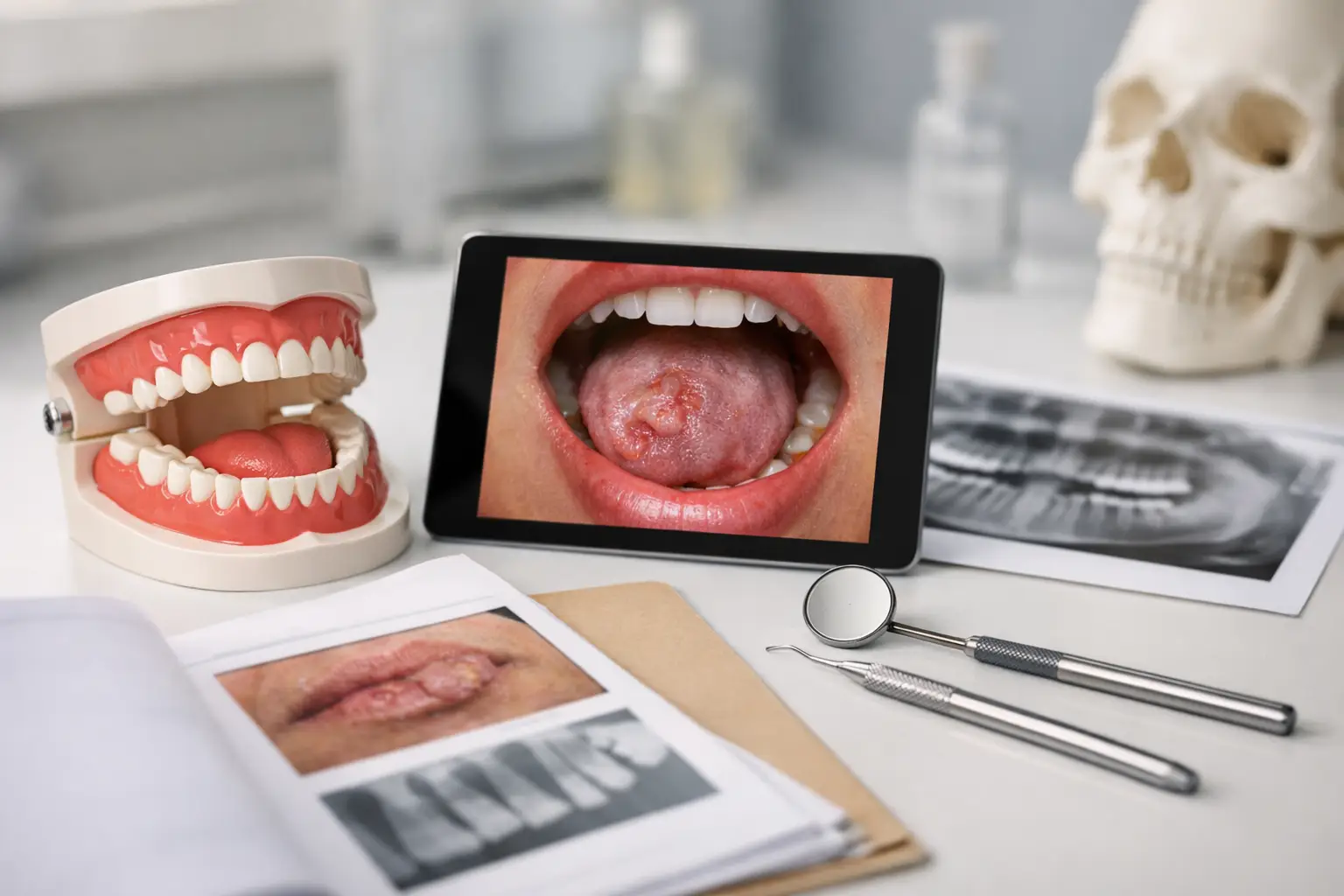
A painless white plaque on the lateral tongue, a patient with burning oral tissues but normal-appearing mucosa, and recurrent ulcers in an otherwise healthy student can all look deceptively straightforward. Oral medicine case studies test whether a candidate can move beyond pattern recognition: identify the relevant history, build a defensible differential diagnosis, recognize urgency, and select the next best step.
For dental students and licensing candidates, the central skill is not memorizing every oral lesion. It is organizing the case so that high-risk findings are not missed and common conditions are not overtreated. This approach applies in US dental curricula and in Canadian undergraduate and national certification settings, even where the examination format and professional regulatory pathway differ.
How to Read Oral Medicine Case Studies
Start by separating the case into five clinical questions: What is the lesion or symptom? Where is it located? How long has it been present? Is it changing? Which host factors alter risk?
Morphology matters, but location and duration often change the level of concern. A frictional keratosis adjacent to a sharp cusp has a different preliminary diagnosis from a non-scrapable white patch on the ventrolateral tongue. Similarly, a single traumatic ulcer that improves after local irritant removal differs from an indurated ulcer persisting for several weeks.
In an examination vignette, do not let a dramatic medical history overshadow the oral findings, or vice versa. A candidate should integrate tobacco and alcohol exposure, immunosuppression, medication use, denture history, systemic symptoms, recent dental treatment, and lesion evolution. The best answer is often the one that addresses the dangerous possibility first, even if a benign diagnosis remains plausible.
A practical reasoning sequence
Use a consistent sequence: describe, localize, characterize, contextualize, and act. Describe whether the finding is white, red, pigmented, ulcerative, vesiculobullous, nodular, or mixed. Localize it to keratinized or nonkeratinized mucosa and note whether it is unilateral, bilateral, focal, or diffuse. Then characterize symptoms, surface texture, scrapability, palpation findings, and duration.
The action step is where many case questions are decided. It may be reassurance and review, elimination of a local cause, laboratory testing, cytology in limited settings, referral, or biopsy. Cytology can support assessment in selected situations, but it does not replace an appropriately planned tissue biopsy when malignancy or a significant immune-mediated disorder is suspected.
Case 1: The Persistent White Lesion
A 58-year-old patient reports a painless white patch on the lateral border of the tongue that has been present for two months. It does not wipe off. The patient smokes cigarettes daily and drinks alcohol regularly. On examination, the lesion is irregular, homogeneous white, and not associated with a sharp tooth or cheek-biting pattern.
The leading clinical concern is leukoplakia, a clinical term for a predominantly white plaque that cannot be characterized as another definable lesion after appropriate evaluation. Leukoplakia is not synonymous with dysplasia or cancer, but it carries malignant potential. The lateral and ventral tongue and floor of mouth are higher-risk sites than many other intraoral locations.
A board-style question may ask for the next step rather than the final histopathologic diagnosis. Observation alone is not the best response when the lesion is persistent, unexplained, and located at a high-risk site in a patient with relevant exposures. Biopsy or prompt specialist referral for biopsy is indicated. If the question instead describes a clear traumatic source and resolution after removing it, reassessment may be appropriate before biopsy.
The key distinction is not simply white lesion versus cancer. It is whether the history and examination adequately explain the lesion, and whether risk features demand tissue diagnosis.
Case 2: White Patches That Wipe Away
A 34-year-old patient using an inhaled corticosteroid for asthma presents with soreness and removable white plaques on the buccal mucosa and dorsal tongue. Under the plaque, the mucosa is erythematous.
This presentation supports pseudomembranous candidiasis. Predisposing factors include inhaled or systemic corticosteroids, broad-spectrum antibiotic exposure, diabetes mellitus, xerostomia, immunosuppression, and denture use. The removable nature of the plaques is a valuable clue, although examination questions occasionally include incomplete descriptions to test whether candidates identify the relevant risk factors.
Management includes addressing the predisposing factor where possible and providing antifungal therapy when clinically indicated. For an inhaled corticosteroid user, rinsing the mouth after use and confirming appropriate inhaler technique can reduce recurrence. A persistent or recurrent infection should prompt consideration of uncontrolled diabetes, immunosuppression, medication-related salivary reduction, or another contributing condition.
Do not confuse candidiasis with leukoplakia merely because both can appear white. Candidiasis is often removable and associated with erythema or soreness; leukoplakia is a non-scrapable clinical diagnosis of exclusion.
Case 3: Recurrent Oral Ulcers
A healthy 22-year-old reports recurrent painful round ulcers on the labial mucosa and floor of mouth. Episodes last 7 to 10 days and recur during periods of stress. There are no skin lesions, ocular symptoms, fever, or gastrointestinal complaints.
This pattern is consistent with recurrent aphthous stomatitis, particularly the minor form when ulcers are small, shallow, and heal without scarring. Aphthae typically affect nonkeratinized mucosa. Recurrent intraoral herpes, by contrast, more commonly affects keratinized mucosa such as attached gingiva and hard palate in immunocompetent patients.
The absence of systemic features supports uncomplicated aphthous disease, but it does not eliminate the need for a careful history. Severe, large, unusually frequent, or persistent ulcers may warrant evaluation for hematinic deficiency, gastrointestinal disease, immune dysfunction, medication effects, or other systemic disease. An examination item may provide recurrent genital ulceration, eye disease, or cutaneous findings to point toward a systemic inflammatory disorder rather than isolated aphthae.
Topical corticosteroids are commonly used to reduce pain and duration in recurrent aphthous stomatitis. The clinical goal is symptom control and recognition of atypical disease, not unnecessary antimicrobial treatment.
Case 4: Burning Mouth With Normal Mucosa
A postmenopausal patient describes daily burning of the anterior tongue and palate, altered taste, and dry-mouth sensation. The oral examination is normal. The patient has no ulceration, erythema, or removable plaques.
Burning mouth syndrome is a diagnosis of exclusion. Before assigning that label, assess for local and systemic causes of oral burning, including candidiasis, xerostomia, nutritional deficiencies, diabetes, thyroid disease, contact reactions, parafunctional habits, and medication effects. A normal examination does not mean that no evaluation is needed.
This case frequently tests an examination principle: symptoms can be genuine and clinically significant even when the mucosa appears normal. The appropriate response is a structured assessment, not dismissal. Management may require patient education, management of contributing factors, symptom-directed therapy, and coordinated medical care when indicated.
Case 5: Desquamative Gingivitis Is a Finding, Not a Diagnosis
A 49-year-old patient presents with painful erythematous gingiva that sloughs during brushing. The patient reports intermittent oral soreness and occasional erosions on the buccal mucosa. Plaque levels are modest and do not explain the severity of inflammation.
Desquamative gingivitis describes the clinical appearance, not the underlying disease. Important causes include mucous membrane pemphigoid, oral lichen planus, pemphigus vulgaris, and contact hypersensitivity reactions. The differential has consequences because some disorders may involve extraoral sites, including ocular tissue, where delayed recognition can be harmful.
A question that includes positive Nikolsky sign, widespread erosions, or ocular symptoms should increase concern for an autoimmune vesiculobullous disorder. Diagnosis typically requires specialist evaluation and biopsy using appropriate technique, often including perilesional tissue for direct immunofluorescence when an immune-mediated blistering disease is suspected. Sampling the center of an ulcerated area may be less diagnostically useful.
What Examiners Commonly Test
Oral medicine questions reward precise distinctions. Candidates should be able to distinguish a clinical descriptor from a definitive diagnosis, a benign reactive lesion from an unexplained persistent lesion, and symptomatic management from treatment of an underlying cause.
They also test prioritization. A persistent ulcer with induration, unexplained erythroplakia, a high-risk white lesion, a rapidly enlarging mass, cervical lymphadenopathy, dysphagia, or unexplained paresthesia should move malignancy higher in the differential and favor urgent referral or biopsy. Erythroplakia is particularly important because it has a greater likelihood of significant epithelial dysplasia or carcinoma on biopsy than many white lesions.
Jurisdictional examination requirements vary. US candidates may encounter integrated biomedical and clinical reasoning through dental school assessments and national board-style examinations, while Canadian candidates may see similar diagnostic principles framed within their undergraduate training and national certification expectations. The science of risk assessment, differential diagnosis, and appropriate referral remains consistent.
A Better Way to Revise Oral Medicine
Instead of studying lesions as isolated flashcards, group them by clinical presentation: non-scrapable white lesions, scrapable white lesions, red lesions, recurrent ulcers, vesiculobullous disease, salivary complaints, and orofacial pain. For each group, learn the discriminating features that change management: site, duration, symptoms, risk factors, palpation, and associated systemic findings.
When reviewing oral medicine case studies, state your reasoning aloud before checking the answer. Name the most likely diagnosis, one serious alternative not to miss, and the next best action. That habit develops the clinical discipline expected in examinations and carries directly into patient care, where the most valuable skill is often recognizing when a familiar-looking finding requires a different response.